Examples of Scans produced by the fluoroscope
The fluoroscopic imaging technique allows us to look at a certain target of the body. It allows us to examine the organ's movement and function. This imaging technique usually used a special radiographic contrast to show contrast between parts. Radiographic contrast shows the different densities in the picture. The dark areas are the tissues that are very dense and the light areas are the less dense. Since the dense tissues absorb more x-ray, it aperars light and the less dense apear dark.This fluoroscopic procedure is usually conducted by a radiologist and an assistant. However, others inside the room while conducting this must wear a protective lead gown and other radiation protective gear.
|
|
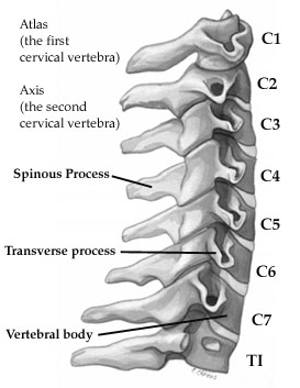
cervical spine
|
Fluoroscopy of the cervical spine
This fluoroscopic image shows the movement of the cervical spine, the spine apears white as because it is the most dense and the other tissues are gray because it is less dense. The colour in this image is taken under low light and so sometimes the bone looks black insted of white. To be more specific of what is happening in this imaging, it shoes the movement of the vertebral body, which is indicated above in the picture at the right. When this part of the spine moved, it lead to the rest also moving because all the parts are attached. However, in the image the vertebral body has the most noticeable movement. Afterwards, it shows the movement of the Spinous process and again all the other parts move but the Spinous process is the most noticeable.
This fluoroscopic image shows the movement of the cervical spine, the spine apears white as because it is the most dense and the other tissues are gray because it is less dense. The colour in this image is taken under low light and so sometimes the bone looks black insted of white. To be more specific of what is happening in this imaging, it shoes the movement of the vertebral body, which is indicated above in the picture at the right. When this part of the spine moved, it lead to the rest also moving because all the parts are attached. However, in the image the vertebral body has the most noticeable movement. Afterwards, it shows the movement of the Spinous process and again all the other parts move but the Spinous process is the most noticeable.
|
Swallowing Disorder
The bones are again the most dense and they are the darkest areas in the imaging and the lightest are other tissues around it that are less dense. In this fluroscopic image, it shows an illness called swallowing disorder and sometimes Dysphagia. The reasons for have a hard time swallowing can be because of Neurological disorders, which is a problem occurring in the nervous system. However, it can also be other reasons such a smoking. In this video, it shows the patient trying to drink liquid. While the liquid is in his/her mouth the epiglottis is closed the oral cavity to prevent food from entering the trachea. However, if a patient did not have a swallowing disorder then, their epiglottis would close naturally without hesitation. It also shows that there is an abnormal contraction in the esophagus. Therefore, this fluoroscopy shows that the patient has a swallowing disorder. |
|
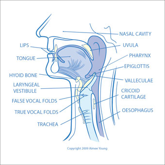
Throat diagram
